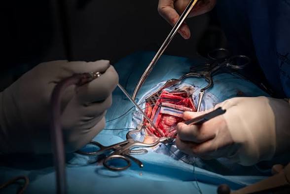
Artificial intelligence has moved from helping doctors analyse scans to assisting them during an actual operation. A 48-year-old British father has become the first known patient to undergo brain surgery with real-time AI guidance, with the technology helping surgeons navigate hidden blood vessels and nerves while removing his tumour.
Highlights
- Rhys Hibbert became the first patient to have brain surgery assisted by live AI guidance.
- Surgeons used the system to identify critical blood vessels and nerves during the operation.
- The tumour was located near the structures responsible for blood supply and vision.
- AI analysed hundreds of previous surgical videos to learn how to recognise key anatomy.
- Doctors say the technology could provide surgeons with an additional expert set of eyes.
Main Story
AI Steps Into the Operating Theatre
A new chapter in medical technology has unfolded in the UK after surgeons successfully used an artificial intelligence system to assist with the removal of a brain tumour while the operation was taking place.
The procedure was carried out at University College London Hospital on Rhys Hibbert, a 48-year-old father of two from Bedfordshire.
Doctors said the tumour was threatening his eyesight because of its position close to the optic nerves.
The experimental AI system analysed a live video feed from the operation and helped surgeons identify areas where important blood vessels and nerves were likely to be located.
A Sudden Health Scare
Hibbert had previously considered himself healthy and active, regularly taking a two-mile lunchtime walk.
That changed when he suddenly collapsed and experienced a seizure while out walking.
Medical scans revealed an 11mm non-cancerous tumour growing on his pituitary gland, a small structure located at the base of the brain.
Although the tumour was not cancerous, its location presented a serious challenge.
The pituitary gland sits close to the carotid arteries, which carry blood to the brain, as well as the optic nerves responsible for vision.
As the tumour grew, it began putting pressure on Hibbert’s optic nerves. He started experiencing reduced peripheral vision, dizziness, severe tiredness and problems with his balance.
Why the Operation Was Risky
Removing a tumour from this part of the brain requires extreme precision.
The surgeons had to work through the nose using an endoscope, a narrow instrument fitted with a camera, before reaching the base of the skull.
The tumour was positioned behind layers of bone and protective tissue, making it difficult to see exactly where critical structures were located.
Doctors also faced the added challenge that every person’s anatomy is slightly different.
According to the information provided by the surgical team, there was a significant possibility that some of the tumour could remain after surgery, while damage to a major blood vessel, although less likely, could have potentially serious consequences.
How the AI Worked
The technology operated on a second screen beside the surgical team.
As the operation progressed, the AI examined the live camera footage, followed the movement of surgical instruments and highlighted areas where important nerves and blood vessels were likely to be found.
Rather than simply showing surgeons an image, the system was trained to recognise anatomical structures that may not be immediately visible.
Researchers developed the technology using hundreds of recordings of previous pituitary tumour operations. Specialists manually identified key blood vessels and nerves in those videos, giving the AI examples from which to learn.
The result was a system capable of recognising patterns in surgical anatomy and providing guidance in real time.
An Extra Set of Eyes for Surgeons
Professor Hani Marcus, one of the specialists involved in the procedure, described the system as an additional expert perspective for surgeons.
The technology is particularly significant because surgeons typically rely heavily on scans taken before an operation to understand a patient’s anatomy.
A real-time system could potentially provide additional information as the procedure unfolds, when the anatomy seen by the surgeon may differ from what was visible on earlier scans.
The researchers say this distinguishes their approach from many other experimental medical AI systems that operate mainly on pre-existing data.
Hibbert Chose to Take Part
For Hibbert, agreeing to participate in the experimental procedure was also about helping advance medicine.
He said the possibility of losing his sight had been frightening, while the illness also affected him emotionally because he did not want to become dependent on others.
Despite those concerns, he chose to take part in the research when doctors offered him the opportunity.
Following the successful removal of the tumour, Hibbert said the experience demonstrated how technology could help surgeons make safer decisions inside an environment where there are no obvious visual markers to guide them.
The operation could offer researchers valuable insight into how artificial intelligence might support surgeons during complex procedures.
The technology is not designed to replace doctors. Instead, its potential lies in giving surgical teams another source of information while they make decisions.
If further testing confirms that the system is safe and reliable, similar technology could eventually be adapted for other highly complex operations.
For patients, the bigger question may no longer be whether AI belongs in healthcare but how far its role in the operating room should go.
As AI gains a place beside surgeons in the operating room, the future of medicine may depend not on replacing human expertise, but on giving it a smarter second pair of eyes.

